 | Info
Sheets |
| | | | | | | | | | | | | | | | | | | | | | | | |
 | Out-
side |
| | | | |
|
| | | | |
Result : Searchterm 'Acquisition Matrix' found in 2 terms [ ] and 10 definitions [ ] and 10 definitions [ ], (+ 5 Boolean[ ], (+ 5 Boolean[ ] results ] results
| | previous 11 - 15 (of 17) nextResult Pages : [1] [2 3] [4] |  | |  | Searchterm 'Acquisition Matrix' was also found in the following service: | | | | |
| |  |
| |
|
| | | | | |  Further Reading: Further Reading: | News & More:
|
|
| |
| | | | | |
| |
|
Quick Overview
Please note that there are different common names for this artifact.
DESCRIPTION
Edge ringing, syrinx-like stripe
A data truncation artifact may occur when the interface between high and low signal intensities is encountered in one imaging plane. The 2D-FT techniques transform the MR signal to spatial intensity image data with frequency and phase information encoding each axis in the plane of the scan. This artifact is found in both frequency and phase axes.
Artifactual ripples adjacent to edges in an image or sharp features in a spectrum, caused by omission of higher frequency terms in Fourier transformation, particularly with the use of zero filling to replace unsampled higher frequencies.
Complex shapes are specified by series of sine and cosine waves of various frequencies, phase and amplitude. Some shapes are more difficult to encode than others. The most difficult shapes to represent with Fourier series of terms are waveforms with instantaneous transitions, tissue discontinuities or edges. The low-frequency components of the series describe the overall shape of the step function. Higher frequency components are needed to describe the corners if the step function more accurately.
If not enough samples are taken, these areas cannot be accurately represented.
The truncation of the infinite data series results in a ringing artifact because of the inability to accurately approximate this tissue discontinuity with a shorter truncated data set. Therefore, the ringing that occurs at all tissue boundaries on MR is called truncation artifact.

Image Guidance
| | | |
• View the DATABASE results for 'Truncation Artifact' (2).
| | | | | | Further Reading: | News & More:
|
|
| |
| | | | |  |
| |
|
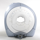
From GE Healthcare;
GE Healthcare has added the Signa HDe 1.5T™, a compact MRI device at an affordable price to its family of MRI products. It has a single electronic cabinet that can be positioned inside the scanner room rather than
in a separate equipment room. The Signa HDe 1.5T can be installed in the same physical location as 0.5T MRI systems with minimal construction costs. According to GE, the installation has been simplified to last only 7 days and has a 30 percent smaller footprint than a typical 1.5T system.
The 1.5T Signa™ HDe MRI system is substantially equivalent to the currently marketed GE 1.5T machines. The data acquisition system supports 1, 4, 8 independent receive channels and multiple independent coil elements per channel during a single acquisition series. The gradient specifications of HDe are lower than other GE Signa 1.5T MRI systems, but it can support clinical applications in cardiac and spectroscopy imaging.
Device Information and Specification CLINICAL APPLICATION Whole body CONFIGURATION Compact short bore 2D 0.7 mm to 20 mm; 3D 0.1 mm to 5 mm 128x512 steps 32 phase encode POWER REQUIREMENTS 480 or 380/415 less than 0.03 L/hr liquid helium | | | |
• View the NEWS results for 'Signa HDe 1.5T™' (1).
| | | | | | Further Reading: | Basics:
|
|
| |
| | | Searchterm 'Acquisition Matrix' was also found in the following service: | | | | |
| | |
| |
|
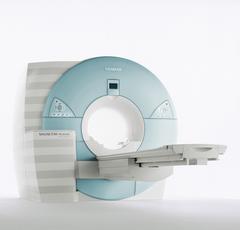
From Siemens Medical Systems;
MAGNETOM Avanto with Tim - Total imaging matrix technology.
For true whole-body anatomical coverage. For ultra-fast image
acquisition. Aids the physician in fast and precise
evaluation of systemic diseases like colon cancer, metastasis imaging, vessel diseases, and preventional exams. For claustrophobic patients,
MAGNETOM Avanto enables feetfirst exams for nearly all MR procedures. For obese patients, MAGNETOM Avanto supports up to 200 kg (400 lbs), without table movement restrictions. The AudioComfort technology enables up to a 30 dB(A) acoustic noise reduction, that means nearly all clinical routine sequences are running under 99 dB(A).
Device Information and Specification
CLINICAL APPLICATION
Whole body
CONFIGURATION
Compact short bore
Body, Tim [32 x 8], Tim [76 x18], Tim [76 coil elements with up to 32 RF channels]
GRE, IR, FIR, STIR, TrueIR/FISP, FSE, FLAIR, MT, SS-FSE, MT-SE, MTC, MSE, EPI, 3D DESS//CISS/PSIF, GMR
IMAGING MODES
Single, multislice, volume study, multi angle, multi oblique
1024 x 1024 full screen display
POWER REQUIREMENTS
380/400/420/440/480 V
Passive, act.; 1st order std./2nd opt.
| | | |
• View the DATABASE results for 'MAGNETOM Avanto™' (2).
| | | | | | Further Reading: | | Basics:
|
|
News & More:
| |
| |
| | | | | |
| |
|
(SCT) The total scan time is the time required to collect all data needed to generate the programmed images.
The scan time is related to the used pulse sequence and dependent on the assemble of parameters like e.g., repetition time (TR), Matrix, number of signal averages ( NSA), TSE- or EPI factor and flip angle.
For example, the total scan time for a standard spin echo or gradient echo sequence
is number of repetitions x the scan time per repetition (means the product of repetition time (TR), number of phase encoding steps, and NSA).
See also Number of Excitations, Turbo Spin Echo Turbo Factor, Echo Planar Imaging Factor, Flip Angle and Image Acquisition Time.
See also acronyms for 'scan time parameters' from different manufacturers. | | | |
• View the DATABASE results for 'Scan Time' (47).
| | |
• View the NEWS results for 'Scan Time' (10).
| | | | | | Further Reading: | Basics:
|
|
News & More:
| |
| |
| | | | |
| | | |
|
| |
 | Look
Ups |
| |


